- India
- International
Thursday, Apr 25, 2024
Journalism of Courage
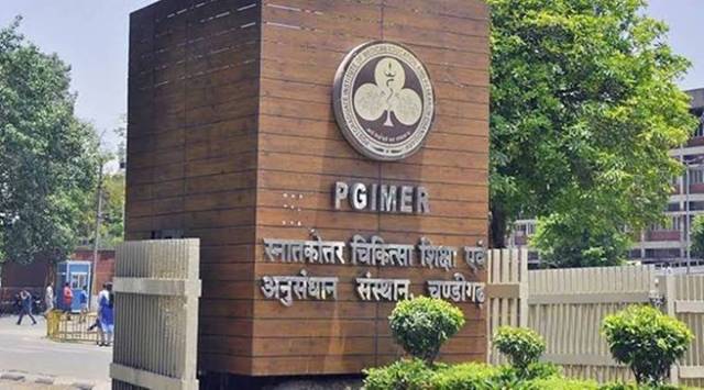 PGIMER's Department of Nephrology has been selected as the Regional Training Centre for South Asia (File)
A team of endoscopic skull base surgeons, Dr Dhandapani SS and Dr Sushant from the Department of Neurosurgery and Dr Rijuneeta from the Department of ENT, PGIMER has created history by operating on the youngest ever patient in the world and removing a large brain tumour (craniopharyngioma), through the nose.
A girl child of one year and four months from Uttarakhand was referred to PGIMER, Chandigarh, with complaints of loss of vision. The child was normal and playful following visual stimuli a few months back. For the last 20 days, the mother noticed that the child was not following anything shown to her. The child’s MRI revealed a calcified brain tumour at the base of the skull, suggestive of craniopharyngioma of size 3 cm, large for a child of one year, close to critical neural structures such as optic nerves and hypothalamus.
These tumours are usually operated on through open surgery, and the remaining part is treated with radiation therapy. Over the last few years, such tumours are being removed through the nose endoscopically by neurosurgeons teaming with ENT surgeons among patients older than six years. However, endoscopic removal through the nose is highly challenging in small children because of small nostrils, immature bones at the skull base, and proximity to crucial blood vessels. The youngest child reported to date having undergone endoscopic surgery through the nose for such tumours was two years old, operated in 2019 at Stanford, USA.
Despite the enormous challenge, Dr Dhandapani chose the endonasal corridor, as the skull opening and brain retraction are avoided if operated through the nose. The team studied the child elaborately using CT angiography navigation and planned for endoscopy. A thin, high- definition endoscope, micro-instruments, and laryngeal coblator were used during the initial steps. Reaching up to the tumour was difficult, as the bones and sinuses were immature. The typical air sinus, which usually gives a corridor to reach up to the tumour base, was absent in this child. The nasal stage was performed by ENT surgeon Dr Rijuneeta, while the skull base part was completed by Neurosurgeons Dr Dhandapani and Dr Sushant.
Extensive drilling of the immature bones was carried out using computer navigation to create a tumour removal corridor. After six hours of surgery, the child was kept in the ICU and recovered well. After ten days, the child has improved vision and no complications, with a CT scan showing almost complete removal.
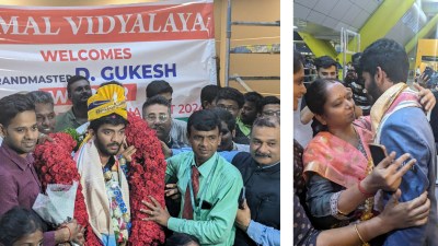
17-year-old D Gukesh, winner of the Candidates chess tournament, received a grand welcome at Chennai airport with a guard of honour from 80 students. Accompanied by his father and coach, he was greeted by officials from AICF and SDAT. His mother, relieved at his return, shared how he rediscovered himself through tennis and socializing.