- News
- City News
- nagpur News
- Maharashtra: Shyam Rathod of Yavatmal carved a niche in the art form called ‘crystal art photomicrography’
Trending
This story is from May 15, 2022
Maharashtra: Shyam Rathod of Yavatmal carved a niche in the art form called ‘crystal art photomicrography’
Shyam Rathod, deputy engineer with MSETCL, has carved a niche for himself in the art form called ‘crystal art photomicrography’.
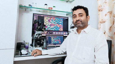
Shyam Rathod, deputy engineer with MSETCL
YAVATMAL: Shyam Rathod, deputy engineer with MSETCL, has carved a niche for himself in the art form called ‘crystal art photomicrography’.
Crystal art photomicrography is a blend of art and science where you passionately photograph art to get beautiful colour textures and microcrystal patterns woven into micro space.
Photographs taken under the polarized microscope are not more than 1x1 mm on the microscope slide.Innovative techniques are used for making a thin layer of crystals on a microscopic slide. The best region on the microscope slide is photographed, which is artistically appealing.
Yavatmal’s Rathod has been pursuing this art form passionately for the past few years. He got to know about this art form from the Facebook group called ‘Crystal Art Photomicrography’. Rathod got an invite from group moderator Loes Modderman of the Netherlands. He also got to know about fellow micro photographers in India like Yogendra Joshi (Pune), Hiroj Bagade (Nagpur) and international professionals.
Rathod experimented with different chemicals to make microcrystals for the slides. This involved either melting chemicals directly or dissolving them in a solvent, then heating and cooling to form crystals. Microcrystals formed start showing beautiful patterns and colours under the microscope. Rathod then experimented with the combination of two or more chemicals like paracetamol and urea, beta alanine and L glutamine etc. The combination of these chemicals yielded crystals.
The crystals, when photographed under the microscope, show beautiful structures and patterns of different colours. For instance, the ABE chemical/medicine used for treating warts, when observed, gave a beautiful flower pattern. Absorbic acid (Vit C) microcrystals formed wave-like structures. The paracetamol and urea combination produced fringe-like structures after many remelts.
Menthol (thandai), available in pan shops, when melted showed different patterns like a heart shape, bubbles and so on.
With the increase in the magnification, the small region of the microcrystal could be observed for intricate details which was magical. No two microcrystals of the same chemical showed identical results even if they were similar. It suggests that time and space, while forming microcrystals, were never the same or identical. The atmospheric conditions and temperature also influence microcrystals.
Crystal art photomicrography is a blend of art and science where you passionately photograph art to get beautiful colour textures and microcrystal patterns woven into micro space.
Photographs taken under the polarized microscope are not more than 1x1 mm on the microscope slide.Innovative techniques are used for making a thin layer of crystals on a microscopic slide. The best region on the microscope slide is photographed, which is artistically appealing.
Yavatmal’s Rathod has been pursuing this art form passionately for the past few years. He got to know about this art form from the Facebook group called ‘Crystal Art Photomicrography’. Rathod got an invite from group moderator Loes Modderman of the Netherlands. He also got to know about fellow micro photographers in India like Yogendra Joshi (Pune), Hiroj Bagade (Nagpur) and international professionals.
The art form requires a polarized microscope, a camera, slides and chemicals to form crystals. It was difficult for Rathod to get these things in a small town like Yavatmal. The cost of a polarized microscope was too high for him. Therefore, he bought a normal compound microscope and converted it into a polarized one by using two polarized filters which he bought online and got them cut into shapes at a local optical shop so that he could fit them in the compound microscope. He also purchased a camera. He was fortunate to get some chemicals from Yogendra Joshi to start with. The next challenge was to connect the camera to the microscope by using a mechanical coupler. The cost of professional couplers was too high and hence he decided to make his own at the local unit. With all the ingredients ready, he was set to embark on the journey of crystal art microphotography.
Rathod experimented with different chemicals to make microcrystals for the slides. This involved either melting chemicals directly or dissolving them in a solvent, then heating and cooling to form crystals. Microcrystals formed start showing beautiful patterns and colours under the microscope. Rathod then experimented with the combination of two or more chemicals like paracetamol and urea, beta alanine and L glutamine etc. The combination of these chemicals yielded crystals.
The crystals, when photographed under the microscope, show beautiful structures and patterns of different colours. For instance, the ABE chemical/medicine used for treating warts, when observed, gave a beautiful flower pattern. Absorbic acid (Vit C) microcrystals formed wave-like structures. The paracetamol and urea combination produced fringe-like structures after many remelts.
Menthol (thandai), available in pan shops, when melted showed different patterns like a heart shape, bubbles and so on.
With the increase in the magnification, the small region of the microcrystal could be observed for intricate details which was magical. No two microcrystals of the same chemical showed identical results even if they were similar. It suggests that time and space, while forming microcrystals, were never the same or identical. The atmospheric conditions and temperature also influence microcrystals.
End of Article
FOLLOW US ON SOCIAL MEDIA
